The beginnings of the Department of Histology and Embryology date back to 1968, when the Laboratory of Histology and Embryology was organized in the Department of Animal Anatomy at a new faculty of the Academy of Agriculture, the Faculty of Veterinary Medicine. The Department of Histology and Embryology was established in 1973 as part of the Institute of Basic Veterinary Sciences. After changes in the faculty structure in 1991, the department was transformed into an independent unit - the Department of Histology and Embryology.
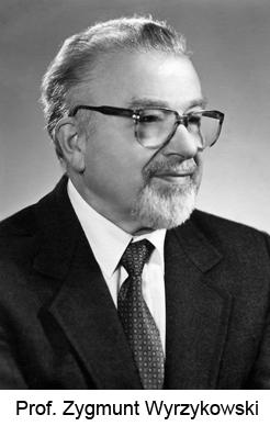
The organizer and first head of the Department in the years 1973 - 1993 was Prof. Zygmunt Wyrzykowski. After that the department was headed by Prof. Dr. Barbara Przybylska-Gornowicz, and from 2010 the Department is headed by Prof. Dr. Bogdan Lewczuk.
 The department was originally located in building 37 in Kortowo I (at 2 Oczapowskiego Street) and from 1985 in building 105 in Kortowo II (at 13 Oczapowskiego Street). Since its foundation, the department has had a histology laboratory and, from 1981, an electron microscopy laboratory. Later, the following laboratories were established: Cell Culture (1992), Radiochemistry (1992) and HPLC (2009). All laboratories have been modernized several times and equipped with increasingly modern equipment. The department also has an animal room where chorobiology experiments can be carried out.
During its existence, the Department of Histology and Embryology has employed three to five people as research and teaching staff and two to three people as technical staff. During its existence, three people have been awarded the title of professor, three people - the title of habilitated doctor and eight people - the title of doctor.
Since 1979, the scientific activities of the Department of Histology and Embryology have focused on research into the morphology and physiology of the pineal gland in mammals and birds. In addition, research is carried out on the influence of various experimental factors on the histology and ultrastructure of the digestive system. Now, the intensively developed research areas concerns the eye structure in health and disease, as well the volume electron microscopy.
The teaching activities of the department were and are mainly focused on the education of the students of the mother faculty in the fields of histology, embryology and cell biology. In addition to the classical teaching methods of these subjects using light and electron microscopes, a virtual microscopy system was introduced in 2009, which significantly improves teaching. The scientific student group of histologists is actively involved in the department's research.